Hypoderma spp. ınvestıgatıon of dynamıc Thıol dısulfıde balance ın ınfested cattle
Abstract
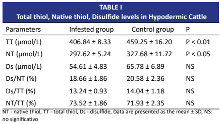
Hipodermiasis in cattle is a parasitic disease caused by Hypoderma bovis and Hypoderma lineatum type fly larvae. The disease, which mostly affects young cattle, causes economic losses such as developmental delay, decrease in meat and milk yield, and inability to use the back skin as a result of being punctured by the larvae. This study aimed to determine serum total thiol, native thiol, disulfide, interleukin-1, IL-6, and tumor necrosis factor-α levels in Hypoderma-infested cattle. In the study, 15 Hypoderma infested cattle diagnosed by clinical examination and 10 healthy cattle for control purposes were used. Serum total thiol, native thiol and disulphide levels of the sick animals were determined as 406.84 ± 8.33 μmol/L, 297.62 ± 5.24 μmol/L, 54.61 ± 4.83 μmol/L, respectively. interleukin-1, interleukin-6 and tumor necrosis factor-α were detected as 41.98 ± 4.37 pg/mL, 94.11 ± 8.49 pg/mL, 82.29 ± 9.12 pg/mL in infested animals. Total thiol, native thiol and disulfide values of infested animals were determined to be lower than the healthy control group, and interleukin-1, interleukin-6, and tumor necrosis factor-α were higher than those of healthy animals. As a result, interleukin-1, interleukin-6, and tumor necrosis factor-α values were determined to be high, and total thiol and native thiol levels were low in cattle with hipodermiasis.
Downloads
References
Dărăbuș G, Tomoioagă V D, Florea T, Imre M, Oprescu I, Morariu S, Mederle N, Ilie MS. Epidemiological surveillance of hypodermosis in cattle from Romania. Pathogens. [Internet]. 2023; 12(9):1077. doi: https://doi.org/q8nh DOI: https://doi.org/10.3390/pathogens12091077
Panadero-Fontán R, Martínez-Calabuig N, García-Dios D. Hypodermosis in Cattle. In: Simões J, ed. Encyclopedia of Livestock Medicine for Large Animal and Poultry Production. Cham, Switzerland: Springer. [Internet]. 2026; p. 634-638. doi: https://doi.org/q8nj DOI: https://doi.org/10.1007/978-3-031-61549-8_49
Zhang H, Dhalla NS. The role of pro-inflammatory cytokines in the pathogenesis of cardiovascular disease. Int. J. Mol. Sci. [Internet]. 2024; 25(2):1082. doi: https://doi.org/gtqww4 DOI: https://doi.org/10.3390/ijms25021082
Pocino K, Carnazzo V, Stefanile A, Basile V, Guerriero C, Marino M, Rigante D, Basile U. Tumor necrosis factor- alpha: Ally and enemy in protean cutaneous sceneries. Int. J. Mol. Sci. [Internet]. 2024; 25(14):7762. doi: https://doi.org/q8nk DOI: https://doi.org/10.3390/ijms25147762
Öztürk G S, Ergun T, Eyüboğlu İ P, Akkiprik M. Serum high- sensitivity C-reactive protein, tumor necrosis factor-α, interleukin (IL)-1β, IL-17A and IL-23 levels in patients with hidradenitis suppurativa. Cytokine. [Internet]. 2021; 144:155585. doi: https://doi.org/gj59cm DOI: https://doi.org/10.1016/j.cyto.2021.155585
Tuzcu N, Tuzcu M, Akçakavak G. Sığırlarda farklı pnömoni tiplerinde tümör nekroz faktör alfa (TNF-α), malondialdehit (MDA), prokalsitonin ve neopterin düzeylerinin karşılaştırılması. Etlik Vet. Mikrobiyol. Derg. [Internet]. 2020; 31(1):52-61. doi: https://doi.org/q8nm DOI: https://doi.org/10.35864/evmd.709433
Iznardo H, Puig L. IL-1 family cytokines in inflammatory dermatoses: pathogenetic role and potential therapeutic implications. Int. J. Mol. Sci. [Internet]. 2022; 23(16):9479. doi: https://doi.org/gq3jcz DOI: https://doi.org/10.3390/ijms23169479
Singh S, Khurana A, Chitkara A. Evaluation of serum levels of interleukins 6, 8, 17 and 22 in acne vulgaris: A cross-sectional study. Indian J. Dermatol. [Internet]. 2023; 68(2):233. doi: https://doi.org/q8nn DOI: https://doi.org/10.4103/ijd.ijd_786_21
Merhan O, Bozukluhan K, Gokce HI. Acute phase proteins and biochemical and oxidative stress parameters in Hypoderma spp. infested cattle. J. Hellenic Vet. Med. Soc. [Internet]. 2017; 68(4):535-540. doi: https://doi.org/q8np DOI: https://doi.org/10.12681/jhvms.16049
Parlak ES, Alisik M, Karalezli A, Sayilir AG, Bastug S, Er M, Hasanoglu HC, Erel O. Are the thiol/disulphide redox status and HDL cholesterol levels associated with pulmonary embolism? Thiol/disulphide redox status in pulmonary embolism. Clin. Biochem. [Internet]. 2017; 50(18):1020-1024. doi: https://doi.org/q8nq DOI: https://doi.org/10.1016/j.clinbiochem.2017.07.018
Merhan O, Taşçı GT, Bozukluhan K, Aydın N. Determination of oxidative stress index and total sialic acid in cattle infested with Hypoderma spp. Kafkas Univ. Vet. Fak. Derg. [Internet]. 2020; 26(5):633-636. doi: https://doi.org/g65pmc
Owegie OC, Kennedy QP, Davizon-Castillo P, Yang M. Thiol Isomerases: Enzymatic Mechanisms, Models of Oxidation, and Antagonism by Galloylated Polyphenols. Antioxidants. [Internet]. 2025; 14(10):1193. doi: https://doi.org/q8ns DOI: https://doi.org/10.3390/antiox14101193
Erel O, Neselioğlu S. A novel and automated assay for thiol/disulphide homeostasis. Clin. Biochem. [Internet]. 2014; 47(18):326–332. doi: https://doi.org/f6r6dk DOI: https://doi.org/10.1016/j.clinbiochem.2014.09.026
Erdoğan H, Camkerten I, Çamkerten G, Ural K, Erdoğan S, Günal İ, Erel Ö. The Effect of hot-iron disbudding on thiol-disulphide homeostasis in calves. Kafkas Univ. Vet . Fak. Derg. [Internet]. 2019; 25(3):335-339. doi: https://doi.org/qzwn
Emre B, Korkmaz O, Koyuncu I, Comakli S, Akcay A, Zonturlu AK, Erel Ö. Determination of thiol/disulphide homeostasis as a new indicator of oxidative stress in dairy cows with subclinical endometritis. Vet . Arhiv. [Internet]. 2021; 91(2):137-148. doi: https://doi.org/q8nt DOI: https://doi.org/10.24099/vet.arhiv.0914
Öktem A, Zenciroğlu A, Öktem A, Bidev D, Özçelik E, Dilli D, Erel Ö. Yenidoğan dönemi pnömoni vakalarında dinamik tiyol/disülfit dengesi. Türkiye Çocuk Hastalıkları Derg. [Internet]. 2021; 15(1):24-29. doi: https://doi.org/q8nv DOI: https://doi.org/10.12956/tchd.746085
Şimşek S, Utuk AE, Köroğlu E, Dumanli N. Türkiye’nin bazı illerindeki sığırlarda hipodermozun seroprevalansı. Res. Vet. Sci. [Internet]. 2008; 84(2):246-249. doi: https://doi.org/d59hjt DOI: https://doi.org/10.1016/j.rvsc.2007.05.007
Ayvazoğlu C, Kiziltepe Ş, Ayvazoğlu-Demir P. Prevalence and economic significance of Hypoderma bovis in Ardahan. S. Afr. J. Anim. Sci. [Internet]. 2022; 52(1):120-125. doi: https://doi.org/q8nw DOI: https://doi.org/10.4314/sajas.v52i1.14
Gingrich RE. Differentiation of resistance in cattle to larval Hypoderma lineatum. Vet. Parasitol. [Internet]. 1980; 7(3):243-254. doi: https://doi.org/b97qtv DOI: https://doi.org/10.1016/0304-4017(80)90028-X
Baron RW, Weintraub J. Lymphocyte responsiveness in cattle previously infested and uninfested with Hypoderma lineatum (de Vill.) and H. bovis (L.) (Diptera: Oestridae). Vet. Parasitol. [Internet]. 1987; 24(3-4):285-296. doi: https://doi.org/bk8fzf DOI: https://doi.org/10.1016/0304-4017(87)90050-1
Panadero R, López C, Parra F, Morrondo P, Díez-Baños P, Colwell DD. Detection of circulating hypodermin C. an antigen capture ELISA for diagnosis of cattle grub (Diptera: Oestridae) infestations. Vet. Parasitol. [Internet]. 2002; 108(1):85–94. doi: https://doi.org/bsjtrg DOI: https://doi.org/10.1016/S0304-4017(02)00179-6
Dacal V, López C, Colwell DD, Vázquez L, Díaz P, Morrondo P, Díez P, Panadero R. Immunohistochemical characterization of inflammatory cells in the skin of cattle undergoing repeated infestations with Hypoderma lineatum (Diptera: Oestridae) larvae. J. Comp. Pathol. [Internet]. 2011; 145(2-3):282-288. doi: https://doi.org/ bn3pkv DOI: https://doi.org/10.1016/j.jcpa.2010.12.015
Uzlu E, Karapehlivan M, Erdoğan HM, Kızıltepe Ş, Erkilıç EE, Deveci HA, Gökçe E, Kaya I, Citil M. Serum and saliva sialic acid and oxidative stress parameters changes in bulls with foot and mouth disease. Kafkas Univ. Vet. Fak. Derg. [Internet]. 2016; 22(3):321-325. doi: https://doi.org/q8nx
Kükürt A, Gelen V, Başer Ö F, Deveci H A, Karapehlivan M. Thiols: Role in oxidative stress-related disorders. In: Atukeren P, ed. Accenting lipid peroxidation. London, United Kingdom: IntechOpen. [Internet]. 2021; 3:96682. doi: https://doi.org/q8nz DOI: https://doi.org/10.5772/intechopen.96682
Circu ML, Aw TY. Reactive oxygen species, cellular redox systems, and apoptosis. Free Radic. Biol. Med. [Internet]. 2010; 48(6):749-762. doi: https://doi.org/dktx2h DOI: https://doi.org/10.1016/j.freeradbiomed.2009.12.022
Espinosa-Diez C, Miguel V, Mennerich D, Kietzmann T, Sánchez-Pérez P, Cadenas S, Lamas S. Antioxidant responses and cellular adjustments to oxidative stress. Redox Biol. [Internet]. 2015; 6:183-197. doi: https://doi.org/f8cv93 DOI: https://doi.org/10.1016/j.redox.2015.07.008
Çetinkaya A. Ratlarda Amiloid Beta1-42 İle Oluşturulan Deneysel Alzheimer Modelinde Tiyol Disülfit Homeostazisi. Düzce Üniv. Sağlık Bil. Enst. Derg. [Internet]. 2020; 10(3):342-347. doi: https://doi.org/q8n2 DOI: https://doi.org/10.33631/duzcesbed.698151
Üstüner P. Vitiligoda anti-oksidan tiyol/disülfit homeostazının rolü: Yeni bir inflamatuvar belirteç. Istanb. Kanuni Sultan Suleym. Tıp Derg. [Internet]. 2018; 10(1):18-24. doi : https://doi.org/q8n3
Değirmençay Ş, Çamkerten G, Çamkerten İ, Aktaş MS. An investigation of thiol/disulfide homeostasis and ischemia- modified albumin levels to assess the oxidative stress in dogs with canine distemper. Vet. Arhiv. [Internet]. 2021; 91(1):39-49. doi: https://doi.org/qzv8 DOI: https://doi.org/10.24099/vet.arhiv.0867
Schmidt EMDS, Fachiolli DF, de Oliveira RM, Almeida FA, Pariz CM, de Lima PR, Costa C, Tvarijonaviciute A, Erel O, Neselioglu S, Ceron JJ, Rubio CP. Changes in serum thiol- disulphide homeostasis in sheep with gastrointestinal nematodes. Animals. [Internet]. 2021; 11(10):2856. doi: https://doi.org/g6svn4 DOI: https://doi.org/10.3390/ani11102856
Aydın Ö, Özkurt G, Çamkerken İ, Eren E, Yanar KE, Aktaş MS. Investigation of Ischemia-Modified Albumin and Thiol/Disulfide Homeostasis for the Determination of Oxidative Stress in sheep with Toxoplasmosis. Small Rumin. Res. [Internet]. 2023; 225:107023. doi: https://doi.org/qzzv DOI: https://doi.org/10.1016/j.smallrumres.2023.107023
Çamkerten İ, Çamkerten G, Erdoğan H, Adnan A, Erdoğan S, Ural K. Serum thiol disulphide levels among sheep with sarcoptic mange. Kafkas Univ. Vet. Fak. Derg. [Internet]. 2019; 25(6):865-868. doi: https://doi.org/q8n4
Deveci MZY, Erdal H. Determination of dynamic thiol- disulfide levels in dairy cattle with foot disease. Vet. Arhiv. [Internet]. 2022; 92(6):657-666. doi: https://doi.org/q8n5 DOI: https://doi.org/10.24099/vet.arhiv.1785
Özcan O, Erdal H, Çakırca G, Yönden Z. Oxidative stress and its impacts on intracellular lipids, proteins and DNA. J. Clin. Exp. Invest. [Internet]. 2015; 6(3):331-336. doi: https://doi.org/p4zh DOI: https://doi.org/10.5799/ahinjs.01.2015.03.0545
Akbaş A, Kilinc F, Sener S, Aktaş A, Baran P, Ergin M. Investigation of thiol-disulphide balance in patients with acute urticaria and chronic spontaneous urticaria. Cutan. Ocul. Toxicol. [Internet]. 2017; 36(3):205-210. doi: https://doi.org/q8n9 DOI: https://doi.org/10.1080/15569527.2016.1240179
Arlian LG, Vyszenski-Moher DL, Rapp CM, Hull BE. Production of IL-1α and IL-1β by human skin equivalents parasitized by Sarcoptes scabiei. J. Parasitol. 1996; 82(5):719-723. doi: https://doi.org/cgzrkz DOI: https://doi.org/10.2307/3283881
Ergönül S, Aşkar TK. Anaplasmosisli sığırlarda ısı şok protein (HSP), Malondialdehit (MDA), nitrik oksit (NO) ve interlökin (IL-6, IL-10) düzeylerinin araştırılması. Kafkas Univ. Vet. Fak. Derg. [Internet]. 2009; 15(4):575-579. doi: https://doi.org/q8pc
Zoroğlan ÇC, Merhan O. Determination of levels of some acute phase proteins, tumor necrosis factor-α, interleukin-1 and interleukin-6 in cattle with trichophythosis. Anim. Health Prod. Hyg. [Internet]. 2023; 12(1):15-19. doi: https://doi.org/q8pf DOI: https://doi.org/10.53913/aduveterinary.1219029
Silva LB, dos Santos-Neto AP, Maia SM, dos Santos- Guimarães C, Quidute IL, Carvalho AD, Júnior SA, Leão JC. The role of TNF-α as a proinflammatory cytokine in pathological processes. Open Dent. J. [Internet]. 2019; 13(1):332-338. doi: https://doi.org/q8ph DOI: https://doi.org/10.2174/1874210601913010332
Hisaeda K, Hagiwara K, Eguchi J, Yamanaka H, Kirisawa R, Iwai H. Interferon-gamma and tumor necrosis factor-α levels in sera and whey of cattle with naturally occurring coliform mastitis. J. Vet. Med. Sci. [Internet]. 2001; 63(9):1009-1011. doi: https://doi.org/d6tppr DOI: https://doi.org/10.1292/jvms.63.1009
Cabanelas E, Panadero R, Baumann A, Alves MP, Summerfield A, García-Dios D, Díaz P, Remesar S, Fernández G, Morrondo MP, Díez-Baños P, López CM. Cytokine expression in bovine PBMC cultures stimulated with Hypoderma lineatum antigens. Vet. Parasitol. [Internet]. 2020; 283:109165. doi: https://doi.org/q8pj DOI: https://doi.org/10.1016/j.vetpar.2020.109165
Vazquez L, Dacal V, Lopez C, Diaz P, Morrondo P, Diez- Banos P, Panadero R. Antigen-specific antibody isotypes, lymphocyte subsets and cytokine profiles in cattle naturally infested by Hypoderma sp.(Diptera: Oestridae). Vet. Parasitol. [Internet]. 2012; 184(2-4):230-237. doi: https://doi.org/b86gdc DOI: https://doi.org/10.1016/j.vetpar.2011.09.013
Ercan N, Tuzcu N, Başbuğ O, Gök K, Işıdan H, Oġrak YZ. The evaluation of important biomarkers in healthy cattle. Kafkas Univ. Vet. Fak. Derg. [Internet]. 2014; 20(5):749- 755. doi: https://doi.org/q8pk DOI: https://doi.org/10.9775/kvfd.2014.11066
Panadero R, López C, Vázquez L, Díaz P, Pérez A, Cabanelas E, Morrondo P, Díez-Baños P. Effect of reinfestations on systemic immune responses in cattle naturally infested by Hypoderma sp.(Diptera: Oestridae). Vet. Parasitol. [Internet]. 2013; 193(1-3):238-244. doi: https://doi.org/q8pm DOI: https://doi.org/10.1016/j.vetpar.2012.11.017