Hepatocutaneous syndrome in a dog with concurrent Ehrlichiosis: Case report
Abstract
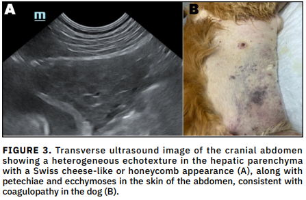
This report describes the diagnosis of hepatocutaneous syndrome in a dog concurrently affected by Ehrlichiosis. A 13-year-old Golden Retriever presented with lethargy, fever, lymphadenopathy, and dermatological lesions on the paws, perianal region, and testes, characterized by hyperkeratosis, erosion, ulceration, crusting, and exudation. Clinicopathological evaluation revealed regenerative anemia, lymphopenia, monocytopenia, basophilia, thrombocytopenia, and increased serum activities of alkaline phosphatase, alanine transaminase, and gamma-glutamyl transferase. After ruling out other vector-borne diseases (e.g., Leishmaniasis, Anaplasmosis, Babesiosis), Ehrlichiosis was diagnosed. Abdominal ultrasonography revealed a honeycomb-like hepatic echotexture, and differential diagnoses for hepatocutaneous syndrome included pemphigus foliaceus/vulgaris, systemic lupus erythematosus, drug eruptions, and zinc-responsive dermatosis. Ultimately, the clinical, laboratory, and ultrasonographic findings supported concurrent diagnoses of hepatocutaneous syndrome and Ehrlichiosis. The dog died, most likely due to severe hepatic dysfunction and associated coagulopathy; disseminated intravascular coagulation was suspected based on the clinical course, although it could not be confirmed due to the absence of coagulation testing, despite receiving combination therapy that included vitamin–amino acid infusion, ursodeoxycholic acid, antioxidant nutraceuticals (S-adenosylmethionine, Silybum marianum, phospholipids, zinc), omega-3 fatty acids, and benzoyl peroxide shampoo for hepatocutaneous syndrome, in addition to doxycycline and prednisolone for ehrlichiosis. This case report highlights the diagnostic complexity of hepatocutaneous syndrome, particularly when concurrent with infectious diseases such as Ehrlichiosis. It demonstrates how overlapping clinical signs can complicate diagnosis and delay appropriate treatment. To the authors’ knowledge, this is the first documented case of hepatocutaneous syndrome in a dog concurrently diagnosed with Ehrlichiosis. By presenting this case, we aim to raise awareness among veterinary clinicians of the importance of a thorough diagnostic workup in dogs exhibiting compatible dermatologic and systemic signs, even in regions endemic for tick- borne diseases.
Downloads
References
Mylonakis ME, Theodorou KN. Canine Monocytic Ehrlichiosis: An Update On Diagnosis and Treatment. Acta Vet. [Internet]. 2017; 67(3):299-317. doi: https://doi.org/gdthgc DOI: https://doi.org/10.1515/acve-2017-0025
Aziz MU, Hussain S, Song B, Ghauri HN, Zeb J, Sparagano OA. Ehrlichiosis in Dogs: A Comprehensive Review about the Pathogen and Its Vectors with Emphasis on South and East Asian Countries. Vet. Sci. [internet]. 2023; 10(1):21. doi: https://doi.org/qrzn DOI: https://doi.org/10.3390/vetsci10010021
Götze DM, Götze MM, Filho ICB, de Carvalho FCG. Superficial Necrolytic Dermatitis in a Dog. Acta Sci. Vet. [Internet]. 2022; 50(supl 1):822. doi: https://doi.org/qrzp DOI: https://doi.org/10.22456/1679-9216.124350
Loftus JP, Rubio MED, Yant J, Bichoupan A, Zhang S, Miller AJ, Center SA, Ruiz MDR, Macho LP. Untargeted metabolomic profiles reveal widespread metabolic perturbations and identify candidate biomarkers in aminoaciduric canine hypoaminoacidemic hepatopathy syndrome. Am. J. Vet. Res. [Internet]. 2023; 84(12):ajvr.23.08.0186. doi: https://doi.org/qrzq DOI: https://doi.org/10.2460/ajvr.23.08.0186
Hall-Fonte DL, Center SA, McDonough SP, Peters-Kennedy J, Trotter TS, Lucy JM, Berger E, Byers C, Cummings CG, Burke E, Stegemen J, Pintar J, Kantrowitz L, Sharpe K, Weinkle T. Hepatocutaneous syndrome in Shih Tzus: 31 cases (1996-2014). J. Am. Vet. Med. Assoc. [Internet]. 2016; 248(7):802-813. doi: https://doi.org/f8f53b DOI: https://doi.org/10.2460/javma.248.7.802
Hill PB, Auxilia ST, Munro E, Genovese L, Silkstone MA, Kirby B. Resolution of skin lesions and long-term survival in a dog with superficial necrolytic dermatitis and liver cirrhosis. J. Small Anim. Pract. [Internet]. 2000; 41(11):519-523. doi: https://doi.org/bzjfgx DOI: https://doi.org/10.1111/j.1748-5827.2000.tb03976.x
Jaffey JA, Backus RC, Sprinkle M, Ruggiero C, Ferguson SH, Shumway K. Successful Long-Term Management of Canine Superficial Necrolytic Dermatitis With Amino Acid Infusions and Nutritionally Balanced Home-Made Diet Modification. Front. Vet. Sci. [Internet]. 2020; 7:28. doi: https://doi.org/qrzv DOI: https://doi.org/10.3389/fvets.2020.00028
Byrne KP. Metabolic epidermal necrosis-hepatocutaneous syndrome. Vet. Clin. North Am. Small Anim. Pract. [Internet]. 1999; 29(6):1337–1355. doi: https://doi.org/qrzw DOI: https://doi.org/10.1016/S0195-5616(99)50131-9
Neer TM, Breitschwerdt EB, Greene RT, Lappin MR. Consensus statement on ehrlichial of small animals from the infectious disease study group of the American College of Veterinary Internal Medicine. J. Vet. Intern. Med. [Internet]. 2002; 16(3):309-315. doi: https://doi.org/c8bn3p DOI: https://doi.org/10.1111/j.1939-1676.2002.tb02374.x
Loftus JP. Aminoaciduric Canine Hypoaminoacidemic Hepatopathy Syndrome. Vet. Clin. North Am. Small Anim. Pract. [Internet]. 2025; 55(4):595-613. doi: https://doi.org/qrzx DOI: https://doi.org/10.1016/j.cvsm.2025.03.009
DeMarle KB, Webster CR, Penninck D, Ferrer L. Approach to the diagnosis of hepatocutaneous syndrome in dogs: a retrospective study and literature review. J. Am. Anim. Hosp. Assoc. [Internet]. 2021; 57(1):15-25. doi: https://doi.org/qrz2 DOI: https://doi.org/10.5326/JAAHA-MS-7072
Outerbridge CA, Marks SL, Rogers QR. Plasma amino acid concentrations in 36 dogs with histologically confirmed superficial necrolytic dermatitis. Vet. Dermatol. [Internet]. 2002; 13(4):177-186. doi: https://doi.org/bm63pq DOI: https://doi.org/10.1046/j.1365-3164.2002.00295.x
Brenseke BM, Belz KM, Saunders GK. Pathology in practice. J. Am. Vet. Med. Assoc. [Internet]. 2011; 238(4):445-447. doi: https://doi.org/d7k2p3 DOI: https://doi.org/10.2460/javma.238.4.445
Leela-arporn R, DeMarle KB, Heinze CR, Webster CRL. Plasma amino acid profiles of dogs with the hepatocutaneous syndrome and dogs with other chronic liver diseases. J. Vet. Intern. Med. [Internet]. 2025; 39(1):e17285. doi: https://doi.org/qrz6 DOI: https://doi.org/10.1111/jvim.17285
Almendros A, Sandy JR, Kirberger RM. Hepatocutaneous syndrome in a Maltese, diagnosis, treatment and the value of CT in the diagnosis. Vet. Rec. Case Rep. [Internet]. 2019; 7(4):e000918. doi: https://doi.org/g4nrf2 DOI: https://doi.org/10.1136/vetreccr-2019-000918
Yoshida M, Barata K, Ando-Lu J, Takahashi M, Maekawa A. A case report of superficial necrolytic dermatitis in a beagle dog with diabetes mellitus. Toxicol. Pathol. [Internet]. 1996; 24(4):498-501. doi: https://doi.org/cx293j DOI: https://doi.org/10.1177/019262339602400413
Isidoro-Ayza M, Lloret A, Bardagi M, Ferrer L, Martínez J. Superficial Necrolytic Dermatitis in a Dog With an Insulin-Producing Pancreatic Islet Cell Carcinoma. Vet. Pathol. [Internet]. 2014; 51(4):805-808. doi: https://doi.org/qrz7 DOI: https://doi.org/10.1177/0300985813503567
Talbot C, Kearns S, Mouser PJ. Treatment of superficial necrolytic dermatitis with copper chelation in a dog with copper-associated hepatitis. J. Am. Anim. Hosp. Assoc. [Internet]. 2023; 59(1):1–6. doi: https://doi.org/qrz8 DOI: https://doi.org/10.5326/JAAHA-MS-7217
Bilgin BH, Kirli PG, Murat H, Turin K. A retrospective epidemiological study: the prevalence of Ehrlichia canis and Babesia volgeli in dogs in the Algean region of Turkey. Acta Vet. [Internet]. 2019; 69(2):164-176. https://doi.org/qrz9 DOI: https://doi.org/10.2478/acve-2019-0013
Qurollo BA, Buch J, Chandrashekar R, Beall MJ, Breitschwerdt EB, Yancey CB, Caudill AH, Comyn A. Clinicopathological findings in 41 dogs (2008-2018) naturally infected with Ehrlichia ewingii. J. Vet. Intern. Med. [Internet]. 2019; 33(2):618–629. doi: https://doi.org/qr2b DOI: https://doi.org/10.1111/jvim.15354
Angkanaporn K, Sanguanwai J, Baiyokvichit TO, Vorrachotvarittorn P, Wongsompong M, Sukhumavasi W. Retrospective analysis of canine monocytic ehrlichiosis in Thailand with emphasis on hematological and ultrasonographic changes. Vet. World. [Internet]. 2022; 15(1):1-9. doi: https://doi.org/pxbg DOI: https://doi.org/10.14202/vetworld.2022.1-9
Mylonakis ME, Kritsepi-Konstantinou M, Dumler JS, Diniz PPVP, Day MJ, Siarkou VI, Breitschwerdt EB, Psychas V, Petanides T, Koutinas AF. Severe Hepatitis Associated with Acute Ehrlichia canis Infection in a Dog. J. Vet. Intern. Med. [Internet]. 2010; 24(3):633–638. doi: https://doi.org/d3xdr3 DOI: https://doi.org/10.1111/j.1939-1676.2010.0501.x
Kaewmongkol G, Lukkana N, Yangtara S, Kaewmongkol S, Thengchaisri N, Sirinarumitr T, Jittapalapong S, Fenwick SG. Association of Ehrlichia canis, Hemotropic Mycoplasma spp. and Anaplasma platys and severe anemia in dogs in Thailand. Vet. Microbiol. [Internet]. 2017; 201:195-200. doi: https://doi.org/f9z89j DOI: https://doi.org/10.1016/j.vetmic.2017.01.022
Loftus JP, Miller AJ, Center SA, Peters-Kennedy J, Astor M. Treatment and outcomes of dogs with hepatocutaneous syndrome or hepatocutaneous-associated hepatopathy. J. Vet. Intern. Med. [Internet]. 2022; 36(1):106-115. doi: https://doi.org/qr2c DOI: https://doi.org/10.1111/jvim.16323