Eficacia comparativa de diferentes fuentes de plasma rico en plaquetas en la cicatrización de heridas cutáneas en conejos diabéticos
Resumen
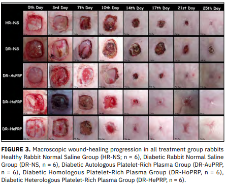
Se evaluaron los efectos terapéuticos del gel de plasma rico en plaquetas de diferentes fuentes en heridas cutáneas de espesor total de 4 cm² en conejos diabéticos. Treinta conejos New Zealand (peso medio: 2.63 ± 0.35 kg; edad: 12–18 semanas) fueron divididos en cinco grupos de seis animales cada uno. El plasma rico en plaquetas heterólogo se obtuvo de una oveja Akkaraman macho (edad: 1 año; peso: 50 kg). Inducción de la diabetes: monohidrato de aloxano (100 mg/kg, 2 dosis, con 48 h de intervalo). Regulación: insulina humana recombinante. Glucemia basal: 100.5 ± 6.5 mg/dL; post-aloxano (5 días): 336.9 ± 51.9 mg/dL (P = 0.000). La doble centrifugación y la gelificación con CaCl₂ produjeron plasma rico en plaquetas con una concentración plaquetaria 3.5 veces superior a la basal. Los parámetros clínicos post-lesión, así como las evaluaciones morfométricas y morfológicas, se realizaron en los días 0, 3, 7, 10, 14, 17, 21 y 25. El análisis histopatológico se realizó el día 25. Se observó una aceleración de la cicatrización en todos los grupos tratados con plasma rico en plaquetas (autólogo diabético-plasma rico en plaquetas, homólogo diabético-plasma rico en plaquetas, heterólogo diabético-plasma rico en plaquetas), mientras que en ambos grupos control se detectó cicatrización retardada, especialmente en el grupo de conejos diabéticos tratados con solución salina. Se observó una mayor contracción de la herida en todos los grupos plasma rico en plaquetas (60 % al día 14) en comparación con los controles (conejo sano con solución salina y diabéticos tratados con solución salina: 56 % en los días 14 y 17, respectivamente). Estos valores alcanzaron posteriormente, en los días 21 y 25, hasta 95.72 ± 3.74 % y 99.23 ± 0.71 % para autólogo diabético-plasma rico en plaquetas; 92.44 ± 5.07 % y 96.04 ± 3.70 % para homólogo diabético-plasma rico en plaquetas; 89.37 ± 12.24 % y 97.32 ± 3.12 % para heterólogo diabético-plasma rico en plaquetas; mientras que fueron 76.96 ± 15.04 % y 84.33 ± 9.29 % para diabéticos tratados con solución salina y 84.91 ± 17.30 % y 92.27 ± 14.72 % para conejo sano con solución salina (P = 0.036 y P = 0.019, respectivamente). Desde el punto de vista histopatológico, el plasma rico en plaquetas potenció la cascada de cicatrización, con angiogénesis temprana, tejido de granulación adecuado y reepitelización suficiente. Independientemente de la fuente de plasma rico en plaquetas, este modula y acelera la cicatrización de heridas cutáneas en modelos de conejos diabéticos, destacando su potencial como una terapia complementaria valiosa para la cicatrización de heridas en pacientes diabéticos.
Descargas
Citas
Abegão KGB, Bracale BN, Delfim IG, Santos ESD, Laposy CB, Nai GA, Giuffrida, RN, Nogueira RMB. Effects of heterologous platelet-rich plasma gel on standardized dermal wound healing in rabbits. Acta Cir. Bras. [Internet]. 2015; 30(3):209-215. doi: https://doi.org/rgzx DOI: https://doi.org/10.1590/S0102-865020150030000008
Baltzis D, Eleftheriadou I, Veves A. Pathogenesis and treatment of impaired wound healing in diabetes mellitus: New insights. Adv. Ther. [Internet]. 2014; 31(8):817-836. doi: https://doi.org/f6f52f DOI: https://doi.org/10.1007/s12325-014-0140-x
Breen A, Mc Redmond G, Dockery P, O'Brien T, Pandit A. Assessment of wound healing in the alloxan-induced diabetic rabbit ear model. J. Investig. Surg. [Internet]. 2008; 21(5):261-269. doi: https://doi.org/dnp94s DOI: https://doi.org/10.1080/08941930802216807
Etulain J. Platelets in wound healing and regenerative medicine. Platelets. [Internet]. 2018; 29(6):556-568. doi: https://doi.org/gh2csg DOI: https://doi.org/10.1080/09537104.2018.1430357
Mihaylova Z, Mitev V, Stanimirov P, Isaeva A, Gateva N, Ishkitiev N. Use of platelet concentrates in oral and maxillofacial surgery: An overview. Acta Odontol. Scand. [Internet]. 2017; 75(1):1-11. doi: https://doi.org/rgzz DOI: https://doi.org/10.1080/00016357.2016.1236985
Gemignani F, Perazzi A, Iacopetti I. Use of canine sourced platelet-rich plasma in a feline contaminated cutaneous wound. Can. Vet. J. [Internet]. 2017 [cited 20 Mar 2026]; 58(2):141-144. PMID: 28216682; PMCID: PMC5234312. Available in: https://goo.su/zrvXtV4
Wang J, Wan R, Mo Y, Zhang Q, Sherwood LC, Chien S. Creating a long-term diabetic rabbit model. Exp. Diabetes Res. [internet]. 2010; 2010:289614. doi: https://doi.org/dmnwf3 DOI: https://doi.org/10.1155/2010/289614
Vendramin FS, Franco D, Franco TR. Método de obtenção do gel de plasma rico emplaquetas autólogo. Rev. Bras. Cir. Plást. [Internet] 2009 [cited 20 Mar 2026]; 24(2):212218. Available in: https://goo.su/fVdteN
Anis A, Sharshar A, El-Hanbally S, Shehata AA. Histopathological evaluation of the healing process of standardized skin burns in rabbits: Assessment of a natural product with honey and essential oils. J. Clin. Med. [Internet]. 2022; 11(21):6417. doi: https://doi.org/rgz2 DOI: https://doi.org/10.3390/jcm11216417
Meira RDO, Braga DNM, Pinheiro LSG, Amorim IFG, Vasconcellos LDS, Alberti LR. Effects of homologous and heterologous rich platelets plasma, compared to poor platelets plasma, on cutaneous healing of rabbits. Acta Cir. Bras. [Internet]. 2020; 35(10):e202001006. doi: https://doi.org/rgz3 DOI: https://doi.org/10.1590/s0102-865020200100000006
Ribeiro G, Martins C, Silva M, Borges V, Lacerda-Neto J. Topical use of kentaserin on healing of experimentallyinduced skin wounds in horses. Arq. Bras. Med. Vet. Zootec. [Internet]. 2009; 61(1):144-148. doi: https://doi.org/dnwhjz DOI: https://doi.org/10.1590/S0102-09352009000100020
Wang J, Wan R, Mo Y, Li M, Zhang Q, Chien S. Intracellular delivery of adenosine triphosphate enhanced healing process in full-thickness skin wounds in diabetic rabbits. Am. J. Surg. [Internet]. 2010; 199(6):823-832. doi: https://doi.org/cd2nk5 DOI: https://doi.org/10.1016/j.amjsurg.2009.05.040
Pradhan L, Nabzdyk C, Andersen ND, LoGerfo FW, Veves A. Inflammation and neuropeptides: The connection in diabetic wound healing. Expert Rev. Mol. Med. [Internet]. 2009; 11:e2. doi: https://doi.org/d2x8f8 DOI: https://doi.org/10.1017/S1462399409000945
Hadour G, Ferrera R, Sebbag L, Forrat R, Delaye J, de Lorgeril M. Improved myocardial tolerance to ischaemia in the diabetic rabbit. J. Mol. Cell. Cardiol. [Internet]. 1998; 30(9):1869-1875. doi: https://doi.org/b6gzd4 DOI: https://doi.org/10.1006/jmcc.1998.0751
Lenzen S. The mechanisms of alloxan- and streptozotocininduced diabetes. Diabetologia. [Internet]. 2008; 51(2):216-226. doi: https://doi.org/dqnhpt DOI: https://doi.org/10.1007/s00125-007-0886-7
Rerup CC. Drugs producing diabetes through damage of the insulin secreting cells. Pharmacol. Rev. [Internet]. 1970; 22(4):485-518. doi: https://doi.org/rg2b DOI: https://doi.org/10.1016/S0031-6997(25)06885-1
Duff GL, Brechin DJ, Finkelstein W. The effect of alloxan diabetes on experimental cholesterol atherosclerosis in the rabbit: IV. The effect of insulin therapy on the inhibition of atherosclerosis in the alloxan - diabetic rabbit. J. Exp. Med. [Internet]. 1954; 100(4):371-380. doi: https://doi.org/c89zz6 DOI: https://doi.org/10.1084/jem.100.4.371
Bell-Jr RH, Hye RJ. Animal models of diabetes mellitus: physiology and pathology. J. Surg. Res. [Internet]. 1983; 35(5):433-460. doi: https://doi.org/b2nrpw DOI: https://doi.org/10.1016/0022-4804(83)90034-3
Spiegelman AR, Tuchman M. Prevention of hypoglycemia during the induction of alloxan diabetes: The use of glucose and antihyaluronidase subcutaneously in the rabbit. Diabetes. [Internet]. 1955; 4(6):473. doi: https://doi.org/rg2c DOI: https://doi.org/10.2337/diab.4.6.473
Froum SJ, Wallace SS, Tarnow DP, Cho SC. Effect of platelet-rich plasma on bone growth and osseointegration in human maxillary sinus grafts: Three bilateral case reports. Int. J. Periodontics Restor. Dent. [Internet]. 2002 [cited 20 Mar 2026]; 22(1):45-53. PMID: 11922217. Available in: https://goo.su/Duf1LwD
Alio JL, Arnalich-Montiel F, Rodriguez AE. The role of “eye platelet rich plasma” (E-PRP) for wound healing in ophthalmology. Curr. Pharm. Biotechnol. [Internet]. 2012; 13(7):1257-1265. doi: https://doi.org/f325f9 DOI: https://doi.org/10.2174/138920112800624355
Kim KM, Shin YT, Kim HK. Effect of autologous plateletrich plasma on persistent corneal epithelial defect after infectious keratitis. Jpn. J. Ophthalmol. [Internet]. 2012; 56:544-550. doi: https://doi.org/f4dwx8 DOI: https://doi.org/10.1007/s10384-012-0175-y
Marx RE. Platelet concentrate: A strategy for accelerating and improving bone regeneration. In: Davies JE, editor. Bone Engineering. Ontario, Toronto, Canada:University of Toronto, Em Squared Inc; 2000. p. 447-453.
Nagata MJ, Messora MR, Furlaneto FA, Fucini SE, Bosco AF, Garcia VG, Deliberador TM, de Melo LG. Effectiveness of two methods for preparation of autologous plateletrich plasma: An experimental study in rabbits. Eur. J. Dent. [Internet]. 2010; 4(04):395-402. doi: https://doi.org/rg2d DOI: https://doi.org/10.1055/s-0039-1697859
Oliveira-Filho MAD, Almeida LE, Pereira JA, Nassif PAN, Czeczko NG, Kume MH, Silva MBG. Platelet-rich plasma in rabbits: introduction of one experimental animal model. Arq. Bras. Cir. Dig. [Internet]. 2008; 21(4):175-179. doi: https://doi.org/d6vj5d DOI: https://doi.org/10.1590/S0102-67202008000400005
Jee CH, Eom NY, Jang HM, Jung HW, Choi ES, Won JH, Hong IH, Kang BT, Jeong DW, Jung DI. Effect of autologous platelet-rich plasma application on cutaneous wound healing in dogs. J. Vet. Sci. [Internet]. 2016; 17(1):79-87. doi: https://doi.org/f8f68h DOI: https://doi.org/10.4142/jvs.2016.17.1.79
DeRossi R, Coelho ACADO, Mello GSD, Frazílio FO, Leal CRB, Facco GG, Brum KB. Effects of platelet-rich plasma gel on skin healing in surgical wound in horses. Acta Cir. Bras. [Internet]. 2009; 24(4):276-281. doi: https://doi.org/d9vsvj DOI: https://doi.org/10.1590/S0102-86502009000400006
Badis D, Omar B. The effectiveness of platelet-rich plasma on the skin wound healing process: A comparative experimental study in sheep. Vet. World. [Internet]. 2018; 11(6):800-808. doi: https://doi.org/gdxjmd DOI: https://doi.org/10.14202/vetworld.2018.800-808
He F, Chen Y, Li J, Lin B, Ouyang Y, Yu B, Xia Y, Ye J. Improving bone repair of femoral and radial defects in rabbit by incorporating PRP into PLGA/CPC composite scaffold with unidirectional pore structure. J. Biomed. Mater. Res. A. [Internet]. 2015; 103(4):1312-1324. doi: https://doi.org/gtd6j3 DOI: https://doi.org/10.1002/jbm.a.35248
Decco OA, Beltrán V, Zuchuat JI, Cura AC, Lezcano MF, Engelke W. Bone augmentation in rabbit tibia using microfixed cobalt-chromium membranes with whole blood and platelet-rich plasma. Materials. [Internet]. 2015; 8(8):4843-4856. doi: https://doi.org/f7q2n9 DOI: https://doi.org/10.3390/ma8084843
Qian Z, Wang H, Bai Y, Wang Y, Tao L, Wei Y, Fan Y, Guo X, Liu H. Improving chronic diabetic wound healing through an injectable and self-healing hydrogel with platelet-rich plasma release. ACS Appl. Mater. Interfaces. [Internet]. 2020; 12(50):55659-55674. doi: https://doi.org/gjps9z DOI: https://doi.org/10.1021/acsami.0c17142
Uraloglu M, Ural A, Efe G, Yulug E, Livaoglu M, Karaçal N. The effect of platelet-rich plasma on the zone of stasis and apoptosis in an experimental burn model. Plast. Surg. [Internet]. 2019; 27(2):173-181. doi: https://doi.org/rg6v DOI: https://doi.org/10.1177/2292550318800498
Acosta L, Castro M, Fernandez M, Oliveres E, Gomez-Demmel E, Tartara L. Treatment of corneal ulcers with platelet rich plasma. Arch. Soc. Esp. Oftalmol. [Internet]. 2014; 89(2):48-52. doi: https://doi.org/f2rzkb DOI: https://doi.org/10.1016/j.oftale.2014.04.012
Schnabel LV, Mohammed HO, Miller BJ, McDermott WG, Jacobson MS, Santangelo KS, Fortier LA. Platelet rich plasma (PRP) enhances anabolic gene expression patterns in flexor digitorum superficialis tendons. J. Orthop. Res. [Internet]. 2007; 25(2):230-240. doi: https://doi.org/dppz2r DOI: https://doi.org/10.1002/jor.20278
Takamura M, Yasuda T, Nakano A, Shima H, Neo M. The effect of platelet-rich plasma on Achilles tendon healing in a rabbit model. Acta Orthop. Traumatol. Turc. [Internet] 2017; 51(1):65-72. doi: https://doi.org/n49d DOI: https://doi.org/10.1016/j.aott.2016.12.001
Roubelakis MG, Trohatou O, Roubelakis A, Mili E, Kalaitzopoulos I, Papazoglou G, Pappa KI, Anagnou NP. Platelet-rich plasma (PRP) promotes fetal mesenchymal stem/stromal cell migration and wound healing process. Stem Cell Rev. Rep. [Internet]. 2014; 10:417-428. doi: https://doi.org/gqd6wq DOI: https://doi.org/10.1007/s12015-013-9494-8
Lee HW, Reddy MS, Geurs N, Palcanis KG, Lemons JE, Rahemtulla FG, Ho KJ, Chen DT, Davis CR, Feldman DS. Efficacy of platelet-rich plasma on wound healing in rabbits. J. Periodontol. [Internet]. 2008; 79(4):691-696. doi: https://doi.org/fxf9rg DOI: https://doi.org/10.1902/jop.2008.070449
Kazakos K, Lyras D, Verettas D, Tilkeridis K, Tryfonidis M. The use of autologous PRP gel as an aid in the management of acute trauma wounds. Injury. [Internet]. 2009; 40(8):801-805. doi: https://doi.org/cf8xdf DOI: https://doi.org/10.1016/j.injury.2008.05.002
Shao S, Pan R, Chen Y. Autologous platelet-rich plasma for diabetic foot ulcer. Trends Endocrinol. Metab. [Internet]. 2020; 31(12):885-890. doi: https://doi.org/gtxnd9 DOI: https://doi.org/10.1016/j.tem.2020.10.003
dos Santos ES, Laposy CB, Abegão KGB, Nogueira RMB, Nai GA, Bracale BN, Delfim IG. Assessment of the healing of standardized wounds in rabbits treated serially with autologous platelet-rich plasma gel. Semina Ciênc. Agrár. [Internet]. 2016; 37(6):4131-4138. doi: https://doi.org/rg6z DOI: https://doi.org/10.5433/1679-0359.2016v37n6p4131
Deng L, Du C, Song P, Chen T, Rui S, Armstrong DG, Deng W. The role of oxidative stress and antioxidants in diabetic wound healing. Oxid. Med. Cell. Longev. [Internet]. 2021; 2021:8852759. doi: https://doi.org/gp4bkd DOI: https://doi.org/10.1155/2021/8852759
Pazzini JM, Nardi ABD, Huppes RR, Gering AP, Ferreira MG, Silveira CP, Luzzi MC, Oliveira JA. Utilização de plasma rico em plaquetas para estimulação da angiogênese em flape de padrão axial toracodorsal em coelhos (Oryctolagus cuniculus). Pesq. Vet. Bras. [Internet]. 2016; 36(2):108118. doi: https://doi.org/rg63 DOI: https://doi.org/10.1590/S0100-736X2016000200008
Kemper B, Brandão CV, Rossetto VJ, Gushiken LF, Padovani CR, Pellizzon CH. Enxertos cutâneos autólogos e homólogos tratados com plasma rico em plaquetas (PRP): Estudo experimental em coelhos. Pesq. Vet. Bras. [Internet]. 2018; 38(9):1818-1823. doi: https://doi.org/rg65 DOI: https://doi.org/10.1590/1678-5150-pvb-5199
Sethi D, Martin KE, Shrotriya S, Brown BL. Systematic literature review evaluating evidence and mechanisms of action for platelet-rich plasma as an antibacterial agent. J. Cardiothorac. Surg. [Internet] 2021; 16(1):277. doi: https://doi.org/rg66 DOI: https://doi.org/10.1186/s13019-021-01652-2
Barrionuevo D, Laposy C, Abegão K, Nogueira R, Nai G, Bracale B, Delfim I. Comparison of experimentallyinduced wounds in rabbits treated with different sources of platelet-rich plasma. Lab. Anim. [Internet] 2015; 49(3):209-214. doi: https://doi.org/f7tz5k DOI: https://doi.org/10.1177/0023677214567747
Carter CA, Jolly DG, Worden-Sr CE, Hendren DG, Kane CJ. Platelet-rich plasma gel promotes differentiation and regeneration during equine wound healing. Exp. Mol. Pathol. [Internet]. 2003; 74(3):244-255. doi: https://doi.org/b846hz DOI: https://doi.org/10.1016/S0014-4800(03)00017-0
Xu P, Wu Y, Zhou L, Yang Z, Zhang X, Hu X, Yang J, Wang M, Wang B, Luo G, He W, Cheng B. Platelet-rich plasma accelerates skin wound healing by promoting reepithelialization. Burns Trauma 2020; 8:tkaa028. doi: https://doi.org/gpk66q DOI: https://doi.org/10.1093/burnst/tkaa028
Hua Y, Bergers G. Tumors vs. chronic wounds: An immune cell's perspective. Front. Immunol. [Internet]. 2019; 10:02178. doi: https://doi.org/gm9c75 DOI: https://doi.org/10.3389/fimmu.2019.02178
Yamada A, Carvalho AM, Oliveira P, Felisbino S, Queiroz D, Watanabe M, Hussni C, Alves ALG. Platelet-rich plasma in the treatment of articular chondral defects experimentally induced in horses: clinical, macroscopic, histopathological and histochemical evaluation. Arq. Bras. Med. Vet. Zootec. [Internet]. 2012; 64(2):323-332. doi: https://doi.org/rg68 DOI: https://doi.org/10.1590/S0102-09352012000200011
Brown DL, Kao WW, Greenhalgh DG. Apoptosis downregulates inflammation under the advancing epithelial wound edge: Delayed patterns in diabetes and improvement with topical growth factors. Surgery. [Internet]. 1997; 121(4):372-380. doi: https://doi.org/bb8pxn DOI: https://doi.org/10.1016/S0039-6060(97)90306-8
Rezende MUD, Silva RBBD, Bassit ACF, Tatsui NH, Sadigursky D, Bolliger-Neto R. Effect of platelet-rich plasma on impact-induced chondrocyte apoptosis. Acta Ortop. Bras. [Internet]. 2011; 19(2):102-105. doi: https://doi.org/rg69 DOI: https://doi.org/10.1590/S1413-78522011000200008
Falanga V. Wound healing and its impairment in the diabetic foot. Lancet. [Internet]. 2005; 366(9498):17361743. doi: https://doi.org/bkz9qd DOI: https://doi.org/10.1016/S0140-6736(05)67700-8
Malavolta EA, Gracitelli MEC, Sunada EE, Benegas E, Prada FdS, Bolliger-Neto R, Rodrigues MB, Ferreira-Neto AA, Camargo OPD. Platelet-rich plasma in arthroscopic repairs of complete tears of the rotator cuff. Rev. Bras. Ortop. [Internet]. 2012; 47(6):741-747. doi: https://doi.org/rg7d DOI: https://doi.org/10.1016/S2255-4971(15)30032-X
Shan GQ, Zhang YN, Ma J, Li YH, Zuo DM, Qiu JI, Cheng B, Chen ZL. Evaluation of the effects of homologous platelet gel on healing lower extremity wounds in patients with diabetes. Int. J. Low. Extrem. Wounds. [Internet]. 2013; 12(1):22-29. doi: https://doi.org/f4qnkf DOI: https://doi.org/10.1177/1534734613477113
Li L, Chen D, Wang C, Yuan N, Wang Y, He L, Yang Y, Chen L, Liu G, Li X, Ran X. Autologous platelet-rich gel for treatment of diabetic chronic refractory cutaneous ulcers: A prospective, randomized clinical trial. Wound Repair Regen. [Internet]. 2015; 23(4):495-505. doi: https://doi.org/f7p9wf DOI: https://doi.org/10.1111/wrr.12294
Elsaid A, El-Said M, Emile S, Youssef M, Khafagy W, Elshobaky A. Randomized controlled trial on autologous platelet-rich plasma versus saline dressing in treatment of non-healing diabetic foot ulcers. World. J. Surg. [Internet]. 2020; 44(4):1294-1301. doi: https://doi.org/rg7c DOI: https://doi.org/10.1007/s00268-019-05316-0
Palumbo VD, Rizzuto S, Damiano G, Fazzotta S, Gottardo A, Mazzola G, Lo Monte AI. Use of platelet concentrate gel in second-intention wound healing: A case report. J. Med. Case Rep. [Internet]. 2021; 15:85. doi: https://doi.org/rg7b DOI: https://doi.org/10.1186/s13256-020-02649-6