Neurotoxicidad Inducida por Cisplatino dependiente de la dosis: Alteraciones histopatológicas y moleculares en el cerebro de rata
Resumen
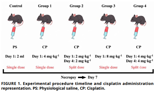
La cisplatina es un agente quimioterapéutico ampliamente utilizado, conocido por causar neurotoxicidad dependiente de la dosis, aunque los mecanismos histopatológicos y moleculares subyacentes aún no se comprenden completamente. El objetivo de este estudio fue investigar los efectos neurotóxicos de diferentes regímenes de dosis de cisplatina en el tejido cerebral de ratas y evaluar las respuestas moleculares asociadas, incluyendo la expresión de factor de necrosis tumoral alfa, proteína de choque térmico 70, péptido natriurético cerebral y subunidad alfa del factor inducible por hipoxia–1. Treinta y ocho ratas albinas Wistar macho se asignaron aleatoriamente a cinco grupos: control, cisplatina en dosis única (4 mg·kg–1 y 8 mg·kg–1) y cisplatina en dosis dividida (2×2 mg·kg–1 y 2×4 mg·kg–1). Tras la administración, se recolectaron tejidos cerebrales para evaluación histopatológica y análisis mediante la reacción en cadena de la polimerasa cuantitativa en tiempo real. Las evaluaciones histopatológicas revelaron necrosis neuronal, gliosis, edema perivascular, neuronofagia y congestión dependientes de la dosis, siendo las alteraciones más graves observadas en el grupo de dosis única de 8 mg·kg–1. Los regímenes de dosis dividida indujeron cambios más leves, sugiriendo que las concentraciones plasmáticas máximas pueden determinar la neurotoxicidad aguda. Los análisis moleculares mostraron una regulación concurrente al alza de factor de necrosis tumoral alfa y proteína de choque térmico 70, indicativa de una respuesta protectora endógena frente a la neuroinflamación inducida por cisplatina. Cabe destacar que los niveles de péptido natriurético cerebral y subunidad alfa del factor inducible por hipoxia–1 mostraron una tendencia al aumento en todos los grupos con neurotoxicidad inducida por cisplatina; estas moléculas no han sido investigadas previamente en el contexto de la neurotoxicidad por cisplatina, lo que resalta un aspecto novedoso de posibles respuestas adaptativas. Los resultados indican que la neurotoxicidad por cisplatina es dependiente de la dosis y más pronunciada con dosis únicas elevadas, mientras que la administración en dosis dividida podría permitir la activación parcial de mecanismos protectores, mitigando el daño histopatológico. En conjunto, estos hallazgos mejoran la comprensión de la lesión cerebral inducida por cisplatina, subrayan la importancia de las estrategias de dosificación clínica para minimizar el riesgo neurotóxico y proporcionan nuevos conocimientos sobre vías moleculares que pueden contribuir a respuestas adaptativas y neuroprotección.
Descargas
Citas
Dos Santos NAG, Ferreira RS, Dos Santos AC. Overview of cisplatin-induced neurotoxicity and ototoxicity, and the protective agents. Food Chem. Toxicol. [Internet]. 2020; 136:111079. doi: https://doi.org/rdm8 DOI: https://doi.org/10.1016/j.fct.2019.111079
Dasari S, Njiki S, Mbemi A, Yedjou CG, Tchounwou PB. Pharmacological effects of cisplatin combination with natural products in cancer chemotherapy. Int. J. Mol. Sci. [Internet]. 2022; 23(3):1532. doi: https://doi.org/gsntwq
Altunkaya M, Ates MB, Bulut A, Abusoglu G, Öztürk B. Cisplatin-induced toxicity in the hippocampus: a dosedependent mechanism of damage. BMC Pharmacol. Toxicol. [Internet]. 2025; 26(1):215. doi: https://doi.org/rdm9
Durak M, Ozhan O, Yildiz A, Durhan M, Vardi N, Cigremis Y, Parlakpinar H. Protective effect of short-term thymoquinone administration on the central nervous system in cisplatininduced neurotoxicity. Eur. Rev. Med. Pharmacol. Sci. [Internet]. 2022; 26(19):6935–6943 doi: https://doi.org/rdnb
Alhowail AH. Cisplatin induces hippocampal neurotoxicity and cognitive impairment in rats through neuroinflammation, oxidative stress, and overexpression of glutamatergic receptors mRNA. Front. Pharmacol. [Internet]. 2025; 16:1592511. doi: https://doi.org/g924xv
Owoeye O, Adedara IA, Farombi EO. Pretreatment with taurine prevented brain injury and exploratory behaviour associated with administration of anticancer drug cisplatin in rats. Biomed. Pharmacother. [Internet]. 2018; 102:375–384. doi: https://doi.org/gndhc9 DOI: https://doi.org/10.1016/j.biopha.2018.03.051
Sekeres MJ, Bradley-Garcia M, Martinez-Canabal A, Winocur G. Chemotherapy-induced cognitive impairment and hippocampal neurogenesis: a review of physiological mechanisms and interventions. Int. J. Mol. Sci. [Internet]. 2021; 22(23):12697. doi: https://doi.org/grrwpd DOI: https://doi.org/10.3390/ijms222312697
George RP, Semendric I, Hutchinson MR, Whittaker AL. Neuroimmune reactivity marker expression in rodent models of chemotherapy-induced cognitive impairment: A systematic scoping review. Brain Behav. Immun. [Internet]. 2021; 94:392–409. doi: https://doi.org/grrwpx DOI: https://doi.org/10.1016/j.bbi.2021.01.021
Basu A, Krishnamurthy S. Cellular responses to Cisplatininduced DNA damage. J. Nucleic Acids [Internet]. 2010; 2010:201367. doi: https://doi.org/c6mnb4 DOI: https://doi.org/10.4061/2010/201367
Yang Z, Schumaker LM, Egorin MJ, Zuhowski EG, Guo Z, Cullen KJ. Cisplatin preferentially binds mitochondrial DNA and voltage-dependent anion channel protein in the mitochondrial membrane of head and neck squamous cell carcinoma: possible role in apoptosis. Clin. Cancer Res. [Internet]. 2006; 12(19):5817–5825. doi: https://doi.org/d7dxft DOI: https://doi.org/10.1158/1078-0432.CCR-06-1037
Hagymasi AT, Dempsey JP, Srivastava PK. Heat–shock proteins. Curr. Protoc. [Internet]. 2022; 2(11):e592. doi: https://doi.org/qk6m DOI: https://doi.org/10.1002/cpz1.592
Becker J, Craig EA. Heat-shock proteins as molecular chaperones. Eur. J. Biochem. [Internet]. 1994; 219(1–2):11– 23. doi: https://doi.org/fxqbzj DOI: https://doi.org/10.1111/j.1432-1033.1994.tb19910.x
Van Noort JM, Bugiani M, Amor S. Heat Shock Proteins: Old and novel roles in neurodegenerative diseases in the Central Nervous System. CNS Neurol. Disord. Drug Targets [Internet]. 2017; 16(3):244–256. doi: https://doi.org/rdnd DOI: https://doi.org/10.2174/1871527315666161031125317
Romi F, Helgeland G, Gilhus NE. Heat-shock proteins in clinical neurology. Eur. Neurol. [Internet]. 2011; 66(2):65–69. doi: https://doi.org/bt6c6j DOI: https://doi.org/10.1159/000329373
Stetler RA, Gan Y, Zhang W, Liou AK, Gao Y, Cao G, Chen J. Heat shock proteins: cellular and molecular mechanisms in the central nervous system. Prog. Neurobiol. [Internet]. 2010; 92(2):184–211. doi: https://doi.org/cngkw5
Hellwig-Bürgel T, Stiehl DP, Wagner AE, Metzen E, Jelkmann W. Review: hypoxia-inducible factor–1 (HIF–1): a novel transcription factor in immune reactions. J. Interferon Cytokine Res. [Internet]. 2005; 25(6):297–310. doi: https://doi.org/c7rtrk DOI: https://doi.org/10.1089/jir.2005.25.297
Basheeruddin M, Qausain S. Hypoxia-Inducible Factor 1–Alpha (HIF–1a): An essential regulator in cellular metabolic control. Cureus [Internet]. 2024; 16(7):e63852. doi: https://doi.org/rdnf DOI: https://doi.org/10.7759/cureus.63852
Chu Q, Gu X, Zheng Q, Zhu H. Regulatory mechanism of HIF–1a and its role in liver diseases: a narrative review. Ann. Transl. Med. [Internet]. 2022; 10(2):109. doi: https://doi.org/rdng DOI: https://doi.org/10.21037/atm-21-4222
Zhang J, Yao M, Xia S, Zeng F, Liu Q. Systematic and comprehensive insights into HIF–1 stabilization under normoxic conditions: implications for cellular adaptation and therapeutic strategies in cancer. Cell. Mol. Biol. Lett. [Internet]. 2025; 30:2. doi: https://doi.org/hbbwkh DOI: https://doi.org/10.1186/s11658-024-00682-7
Schumacker PT. Hypoxia-inducible factor–1 (HIF–1). Crit. Care Med. [Internet]. 2005; 33(12):423–425. doi: https:// doi.org/ffg57v DOI: https://doi.org/10.1097/01.CCM.0000191716.38566.E0
Howell NJ, Tennant DA. The role of HIFs in ischemia-reperfusion injury. Hypoxia [Internet]. 2014; 2:107–115. doi: https://doi.org/qk6k DOI: https://doi.org/10.2147/HP.S49720
Zimna A, Kurpisz M. Hypoxia-Inducible Factor–1 in physiological and pathophysiological angiogenesis: applications and therapies. Biomed. Res. Int. [Internet]. 2015; 2015:549412. doi: https://doi.org/gb57nx DOI: https://doi.org/10.1155/2015/549412
Balamurugan K. HIF–1 at the crossroads of hypoxia, inflammation, and cancer. Int. J. Cancer [Internet]. 2016; 138(5):1058–1066. doi: https://doi.org/ggwwk2 DOI: https://doi.org/10.1002/ijc.29519
Nakagawa Y, Nishikimi T, Kuwahara K. Atrial and brain natriuretic peptides: Hormones secreted from the heart. Peptides [Internet]. 2019; 111:18–25. doi: https://doi.org/rdnj DOI: https://doi.org/10.1016/j.peptides.2018.05.012
Alcidi G, Goffredo G, Correale M, Brunetti ND, Iacoviello M. Brain natriuretic peptide biomarkers in current clinical and therapeutic scenarios of heart failure. J. Clin. Med. [Internet]. 2022; 11(11):3192. doi: https://doi.org/g83qkw DOI: https://doi.org/10.3390/jcm11113192
Katoh C, Osanai T, Tomita H, Okumura K. Brain natriuretic peptide is released from human astrocytoma cell line U373MG under hypoxia: a possible role in anti-apoptosis. J. Endocrinol. [Internet]. 2011; 208(1):51–57. doi: https://doi.org/bz44g9 DOI: https://doi.org/10.1677/JOE-10-0230
Panagopoulou V, Deftereos S, Kossyvakis C, Raisakis K, Giannopoulos G, Bouras G, Pyrgakis V, Cleman MW. NTproBNP: an important biomarker in cardiac diseases. Curr. Top. Med. Chem. [Internet]. 2013; 13(2):82–94. doi: https:// doi.org/f4rsds DOI: https://doi.org/10.2174/1568026611313020002
Volarevic V, Djokovic B, Jankovic MG, Harrell CR, Fellabaum C, Djonov V, Arsenijevic N. Molecular mechanisms of cisplatininduced nephrotoxicity: a balance on the knife edge between renoprotection and tumor toxicity. J. Biomed. Sci. [Internet]. 2019; 26(1):25. doi: https://doi.org/gqf7z3 DOI: https://doi.org/10.1186/s12929-019-0518-9
Zavala-Valencia AC, Velasco-Hidalgo L, Martínez-Avalos A, Castillejos-López M, Torres-Espíndola LM. Effect of N-Acetylcysteine on cisplatin toxicity: a review of the literature. Biol.: Targets Ther. [Internet]. 2024; 18:7–19. doi: https://doi.org/hbbpjw DOI: https://doi.org/10.2147/BTT.S438150
Aksit D, Yazici A, Aksit H, Sari ES, Yay A, Yildiz O, Kiliç A, Ermis SS, Seyrek K. Selenium protects retinal cells from cisplatininduced alterations in carbohydrate residues. Balkan Med. J. [Internet]. 2016; 33(4):441–447. doi: https://doi.org/rdnk DOI: https://doi.org/10.5152/balkanmedj.2015.155532
Nachnani R, Sepulveda DE, Booth JL, Zhou S, Graziane NM, Raup-Konsavage WM, Vrana KE. Chronic cannabigerol as an effective therapeutic for cisplatin-induced neuropathic pain. Pharmaceuticals [Internet]. 2023; 16(10):1442. doi: https://doi.org/g9853f DOI: https://doi.org/10.3390/ph16101442
Karavelioglu E, Boyaci MG, Simsek N, Sonmez MA, Koc R, Karademir M, Guven M, Eser O. Selenium protects cerebral cells by cisplatin induced neurotoxicity. Acta Cir. Bras. [Internet]. 2015; 30(6):394–400. doi: https://doi.org/rdnn DOI: https://doi.org/10.1590/S0102-865020150060000004
Kazak F, Akalin PP, Yarim GF, Baspinar N, Özdemir Ö, Ates MB, Altug ME, Deveci MZY. Protective effects of nobiletin on cisplatin induced neurotoxicity in rats. Int. J. Neurosci. [Internet]. 2022; 132(5):531–537. doi: https://doi.org/rdnp
Abdel-Wahab WM, Moussa FI. Neuroprotective effect of N-acetylcysteine against cisplatin-induced toxicity in rat brain by modulation of oxidative stress and inflammation. Drug Des. Dev. Ther. [Internet]. 2019; 13:1155–1162. doi: https://doi.org/rdnq DOI: https://doi.org/10.2147/DDDT.S191240
Gutte H, Oxbøl J, Kristoffersen US, Mortensen J, Kjær A. Gene expression of ANP, BNP and ET–1 in the heart of rats during pulmonary embolism. PLoS One [Internet]. 2010; 5(6):e11111. doi: https://doi.org/dnnmwb DOI: https://doi.org/10.1371/journal.pone.0011111
Wang X, Li X, Erhardt JA, Barone FC, Feuerstein GZ. Detection of tumor necrosis factor–a mRNA induction in ischemic brain tolerance by means of real-time polymerase chain reaction. J. Cereb. Blood Flow Metab. [Internet]. 2000; 20(1):15–20. doi: https://doi.org/bhs6nw DOI: https://doi.org/10.1097/00004647-200001000-00004
Bulut A, Tuzcu M, Ates MB, Çetin SH, Kaya V. Análisis histopatológico y PCR en tiempo real de alteraciones en el cerebro de rata en el síndrome de aplastamiento inducido por glicerol. Rev. Cient. FCV-LUZ [Internet]. 2026; 36(1):e361779. doi: https://doi.org/rdns
Elghamry HA, Mohamed MI, Hassan FM, Abdelfattah DS, Abdelaal AG. Potential use of GAPDH m-RNA in estimating PMI in brain tissue of albino rats at different environmental conditions. Egypt. J. Forensic Sci. [Internet]. 2017; 7(1):24. doi: https://doi.org/ghj4s3 DOI: https://doi.org/10.1186/s41935-017-0024-8
Moneim AEA. Azadirachta indica attenuates cisplatin-induced neurotoxicity in rats. Indian J. Pharmacol. [Internet]. 2014; 46(3):316–321. doi: https://doi.org/rdnt DOI: https://doi.org/10.4103/0253-7613.132182
Öztopuz RÖ. Melatonin ameliorates cisplatin-induced neurodegeneration in medulla oblongata through the expressions of Aqp–1,–4, inflammation and apoptosis pathway genes. Turk. J. Biol. [Internet]. 2022 [cited 20 Mar 2026]; 46(2):162–172. Available in: https://goo.su/qASoK DOI: https://doi.org/10.55730/1300-0152.2583
Khadrawy YA, El-Gizawy MM, Sorour SM, Sawie HG, Hosny EN. Effect of curcumin nanoparticles on the cisplatin-induced neurotoxicity in rat. Drug Chem. Toxicol. [Internet]. 2019; 42(2):194–202. doi: https://doi.org/rdnv DOI: https://doi.org/10.1080/01480545.2018.1504058
Oliveira HR, Coelho MS, Neves FAR, Duarte DB. Cisplatininduced changes in calcitonin gene-related peptide or TNF–a release in rat dorsal root ganglia in vitro model of neurotoxicity are not reverted by rosiglitazone. Neurotoxicology [Internet]. 2022; 93:211–221. doi: https://doi.org/rdnw DOI: https://doi.org/10.1016/j.neuro.2022.10.002
Michel L, Rassaf T, Totzeck M. Biomarkers for the detection of apparent and subclinical cancer therapy-related cardiotoxicity. J. Thorac. Dis. [Internet]. 2018; 10(35):4282–4295. doi: https://doi.org/gnb6r7 DOI: https://doi.org/10.21037/jtd.2018.08.15
Chang P, Zhang X, Zhang J, Wang J, Wang X, Li M, Wang R, Yu J, Fu F. BNP protects against diabetic cardiomyopathy by promoting Opa1–mediated mitochondrial fusion via activating the PKG-STAT3 pathway. Redox Biol. [Internet]. 2023; 62:102702. doi: https://doi.org/qk6v DOI: https://doi.org/10.1016/j.redox.2023.102702
Van Molle W, Wielockx B, Mahieu T, Takada M, Taniguchi T, Sekikawa K, Libert C. HSP70 protects against TNF-induced lethal inflammatory shock. Immunity [Internet]. 2002; 16(5):685–695. doi: https://doi.org/ccq65s DOI: https://doi.org/10.1016/S1074-7613(02)00310-2
Belenichev IF, Aliyeva OG, Popazova OO, Bukhtiyarova NV. Involvement of heat shock proteins HSP70 in the mechanisms of endogenous neuroprotection: the prospect of using HSP70 modulators. Front. Cell. Neurosci. [Internet]. 2023; 17:1131683. doi: https://doi.org/qk6w